Egg Anatomy
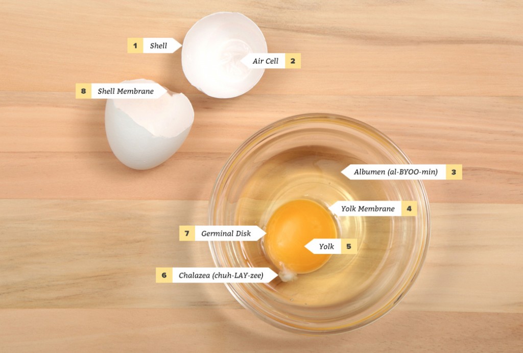
- Shell: The shell, which contains approximately 10,000 tiny pores that allow moisture and gasses in and out, is the egg’s first line of defense against the entry of bacteria.
- Air Cell: An air cell is formed at the wide end of the egg as it cools off after being laid. The fresher the egg, the smaller the air cell.
- Albumen: Albumen is the egg white, which represents 2/3 of an egg’s weight. There are two layers – thick and thin albumen – that are made mostly of water, high-quality protein and minerals.
- Yolk Membrane (vitelline membrane): The yolk membrane surrounds and holds the yolk. The fresher the egg, the stronger the yolk membrane.
- Yolk: The yolk is the egg’s major source of vitamins and minerals, which represents 1/3 of an egg’s weight. The yolk colour can range from light yellow to dark orange, depending on the hen’s feed.
- Chalazea: Chalazea is a pair of spiral bands that anchor the yolk in the centre of the albumen. The fresher the egg, the more prominent the chalazea.
- Germinal Disk: The germinal disk appears as a slight depression of the surface of the yolk, which is the entry for the fertilization of the egg.
- Shell Membrane: There are two membranes on the inside of the shell (one sticks to the shell and the other surrounds the albumen), which together are the egg’s second line of defense against bacteria.